Many, if not all, of us know someone who is battling cancer or has battled this type of cancer. According to the National Cancer Institute, one in eight women who live to be 80 years old will develop breast cancer. It is the second most commonly diagnosed cancer among women, just after skin cancer. While most breast cancer diagnoses are for women, a number of them are diagnosed in men. The breast is composed of glands that make breast milk known as lobules, small tubes that carry the milk from the lobules to the nipples known as ducts, tissue, and blood and lymph vessels. The two areas that are most likely to develop cancerous cells are the ducts and glands, but in rarer cases it can develop in the other areas. Breast cancer may also develop from lymph nodes that surround the breast, like those of the underarm.
The progression of cancer is the result from a series of changes, which include the inactivation of tumor suppressor genes and the activation of oncogenes. There are different oncogenes that are discussed in the Molecular basis of breast cancer that are linked to breast tumor progression. These include p53, BCL-2, Bag-1, P27, Skp2, HER-2, and the estrogen receptors (ER). P53 is a tumor suppressor gene and is involved in the regulation of normal cell growth and division. Disruption of the function of p53 results in continued survival allowing continued growth of damaged cells. Bcl-2 is a family of genes that can either be pro-survival or pro-apoptotic (death of cells). A balance between these different types controls the development and function of normal breasts. Bag-1 is a Bcl-2 interacting anti-apoptotic protein. It also binds hormone receptors and inhibits hormone induced cell death. Bag-1 plays a key role in the oncogenesis and progression of breast cancer. Lowered expression of p27 is linked to overall survival and shorter time to progression. Loss of p27 is linked to the change of cells from normal to premalignant to malignant. S-phase kinase-association protein, or Skp2, is needed for degradation of p27. HER-2 is a promising tumor marker. Evidence shows that over-expression of HER-2 is involved in the progression of breast cancer. Another molecular marker association with breast cancer is the estrogen receptor (ER), which plays a key role in the rapid increase of responsive cells.
References:
http://www.cancercenter.com/breast-cancer/?source=GGLPS01&channel=paid+search&invsrc=Non_Branded_Paid_Search_Google_Cancer_Search&utm_device=c&utm_budget=Corporate&utm_site=GOOGLE&utm_campaign=Non+Brand%3ECancer+Type%3A+Breast&utm_adgroup=Breast+Cancer%3EGeneral%3EExact&utm_term=breast+cancer&utm_matchtype=e&k_clickid=9f30dc28-88bc-4c7f-9ebf-59cb923353f4&k_profid=422&k_kwid=3852244
http://smj.org.sa/index.php/smj/article/viewFile/3481/1255
Monday, December 5, 2016
Friday, November 18, 2016
Melanoma
This weeks cancer topic is melanoma skin cancer. Melanoma is a cancer that begins in the skin cells. Melanoma begins specifically in the melanocyte type of skin cells. Melanocytes make a brown pigment known as melanin, which is what gives skin the brown or tan color. When you lay out to tan and are exposed to more sunlight the melanocytes will make more of the pigment. Melanomas can occur anywhere on your skin, but primarily occur on the chest or back in men and the legs of women. This type of skin cancer can come from moles that turn into melanoma. UV rays from sunlight or from tanning beds are a major cause of melanoma. These UV rays damage the DNA in the skin, which can alter how genes control how skin cells grow and divide. DNA changes can turn on oncogenes, which help cells grow and divide, or turn off tumor suppressor genes, which keep cell growth in check or cause cells to die.
Current research suggests that increased sun exposure as a child and teenager is linked to development of melanoma. Majority of those with melanoma have reported having sunburns or other intense sun exposures as a child. Researcher have also noted that their has been an increase in melanomas occurring on the arms, neck, and face. These areas are commonly exposed, especially in men, which could lead to a higher risk of melanoma. Researchers are studying whether or not these different types of exposures to UV (including tanning booths) creates changes in different genes. If this is the case, different treatments may be needed for different exposure histories.
While it is scary to think that our exposure to sun as a child and teenager could be the reason for developing melanoma later in life, we should still take precautions to avoid getting skin cancer. The best thing you can do is wear sunscreen, limit your exposure to UV rays, and know your body. If you notice any strange discoloration, growths, bumps, patches, or sores that won't go away.
Reference:
http://www.cancer.org/cancer/skincancer-melanoma/
Wednesday, November 9, 2016
Hodgkin Lymphoma
Hodgkin's lymphoma is a type of lymphoma, which is a group of blood cell tumors that develop from lymphocytes. Generally one will experience fevers, night sweats, and weight loss. A proportion of Hodgkin's tumors contain the Epstein-Barr virus (EBV), which suggests that this virus is a contributing factor to the develop of the cancer. EBV, or human herpesvirus 4, is one of the known viruses in the herpes family. The virus infects the B cells of the immune system and epithelial cells, and once the infection is under control EBV latency will persist for the rest of the individual's life.
But how does this virus lead to Hodgkin's lymphoma? The mechanism by which EBV might cause Hodgkin's lymphoma is not well understood. However, almost 40% of individuals who have Hodgkin's lymphoma are also infected with EBV. EBV possibly triggers the pathogenic mechanism of Hodgkin's disease, which means that EBV could play a direct or indirect role in he pathogenesis. According to Flavell, cloned viral probes and Southern blot methods were used to detect EBV DNA in 20-25% of Hodgkin's disease tumor specimens. This approach, however, could not determine the locality of the EBV genome in the tissues. This method of detecting EBV DNA was the first to demonstrate its existence in Hodgkin-Reed Sternberg (HRS) cells. HRS cells are different large cells found in individuals with Hodgkin's lymphoma.
Those that are diagnosed with Hodgkin's lymphoma will generally undergo radiation therapy or chemotherapy. Sometimes both treatments are used, but this depends on how far the disease has progressed in the individual. Luckily, there is a high cure rate and long survival for patients battling this disease.
Link to the paper used:
https://www.ncbi.nlm.nih.gov/pmc/articles/PMC1186979/
Friday, November 4, 2016
HPV in Men
Continuing off of my previous blog, this week I will be discussing human papillomavirus (HPV) and its role in males. In case you missed my previous blog, HPV is a common infection to those who are sexually active. However, most infections will go away on their own without any further problems. Getting the HPV vaccine can help prevent the onset of cervical cancer or genital warts that may occur from HPV. This vaccine is very common for teenage girls to receive, but should males be receiving this vaccine too?
The answer is yes, but there are some considerations. According to the CDC, it is recommended that boys between the ages of 11 and 12 receive the vaccine, but boys as young as 9 and through the age of 21 can receive the vaccine. While HPV isn't cancer, it causes certain changes in the body that can lead to cancer. While cervical cancer is what we hear about most, there are cancers that can occur in males from this infection. Males can develop penile cancer if the infection doesn't go away. Both men and women can develop anal cancer and even cancer in the back of the throat, known as oropharyngeal cancer. Good news, however, is that HPV-related cancers are not very common in men even though HPV is one of the most commonly transmitted infections. Not so good news is that there are currently no approved tests for HPV in men and there are also no specific treatments for HPV. There are only treatments for the symptoms, such as genital warts.
Most of the cancers caused by HPV can be prevented with the HPV vaccination. Approximately 63% of penile cancers are linked to HPV, along with 91% for anal cancers, and 72% for cancer of the back of the throat. So how can HPV lead to cancer? HPV is a DNA virus that contains genes which encode for biological processes that allow the virus to in the cell nucleus of its' host cell. The virus will replicate in the nucleus until cell death. At this point, the virus will then be allowed to infect other epithelial cells and eventually obtain the ability to embed itself into the DNA of the host. This gives the virus the ability to control cellular mechanisms and will mess up cell regulation. This deregulation will cause uncontrolled cell growth, which in turn causes the cancers that have been discussed. While not all HPV types have this ability, as discussed in my previous blog, there are certain cancer-causing types that could occur.
For more information from my readings here are the links:
http://www.cdc.gov/std/hpv/stdfact-hpv-and-men.htm
http://www.canceradvocacy.ca/reportcard/2012/Report%20Card%20on%20Cancer%20in%20Canada%202011-12.pdf#page=6 (page 6 "HPV in Men")
Friday, October 28, 2016
The molecular basis of human papillomavirus
Around the age of 13, I had to get the vaccine for the human papillomavirus (HPV), as I'm sure most females have. I have never looked into what this virus is and why I had to receive this vaccine. I started my search with Molecular biology of human papillomavirus infection and cervical cancer. It turns out, there are over 100 different HPV types that have been identified. These different types of HPVs cause a variety of epithelial lesions. The two main HPVs are the alpha and beta papillomaviruses. Beta papillomaviruses can spread if unchecked and can lead to the development of non-melanoma skin cancer. Alpha papillomavirus is the largest group of HPVs out of all of the types.
Alpha papillomaviruses cause warts, which are usually not linked to cancer in most patients. However, there are 30 types of HPVs that infect the cervical epithelium and part of these cause lesions that can lead to cancer and are labeled as "high-risk" HPV. The most common high-risk HPV type is known as HPV16, which causes about 50% of cervical cancers. High-risk HPV infections occur in younger woman (about 25), approximately 20-40%. As you get older, however, the risk of cervical cancer is lowered due to the infections clearing up or the immune system gaining control of the infection.
Human papillomaviruses are circular with double stranded DNA and have a protective capsid. This virus will infect the epithelial cells of the cervical mucosa and integrates the DNA into the cellular genome. While 90% will heal within approximately two years, some instances involve the HPV DNA integrating into tumor cell DNA and developing into cancer. The life cycle of HPV includes the initial infection, uncoating, amplification of its' genome, and packaging to form new viral particles. The papillomaviruses will cause cancer when regulation of gene expression is disturbed. Oncoproteins E6 and E7 are apart of the regulation of cell proliferation and cell death. When these are expressed incorrectly it can be extremely dangerous and leads to the cancer causing papillomavirus.
Reference:
http://www.clinsci.org/content/110/5/525.full
Friday, October 21, 2016
Lydia Villa-Komaroff

Dr. Villa-Komaroff was apart of a research team the worked on cloning insulin in 1977. She was successfully apart of the group that conducted the first synthesis of mammalian insulin in bacterial cells with the use of recombinant DNA. Her research also includes the discovery of a molecule associated with Alzheimer's disease causes degeneration of neurons in the brain. This molecule is known as amyloid beta, and before her research it was not very clear if this molecule produced the degeneration or was a byproduct of the degeneration of the neurons. This discovery led to further research on treating Alzheimer's by targeting the amyloid beta molecule.
If her groundbreaking research wasn't amazing enough, Dr. Villa-Komaroff has attained numerous awards and honors. A number of her awards have been in regards for Hispanics or women in science. This just goes to show that she was able to break through the barriers she faced in her early scientific career.
Here is an article on her:
http://www.unhny.org/programs-and-issues/stem-spotlight-lydia
Here is her article regarding her research on cloning insulin:
http://www.pnas.org/content/75/8/3727.full.pdf
Friday, October 14, 2016
Don't FRET!
Fluorescence Resonance Energy Transfer, that is. This technique is used for probing intermolecular interactions and determining spatial measurements of macromolecules. I recently heard about this technique for the first time and it sparked my interest.
FRET utilizes chromophores, which is the part of the molecule that produces color through absorption and transmittance. The donor chromophore, which is in an excited state, will transfer its energy non-radiatively to the acceptor chromophore. At a certain wavelength, the acceptor chromophore will absorb in the same region that the donor chromophore will emit fluorescence. When the two chromophores are far enough apart, you will only see the donor chromophore lit up. When they get closer together, the donor will give it's energy to the acceptor chromophore, allowing the acceptor to light up and the donor to not. The donor will only transfer the energy to the acceptor when they are in close proximity to each other. Since this energy transfer only occurs at certain distances, there are a variety of uses for this technique.
FRET can be used to examine intermembrane exchange of lipid components, vesicle-vesicle interactions, and membrane fusion, as these all involve changes in distances. It has been used to look at viruses and how they fuse to cells. Interactions between membrane proteins and the components of the membrane in cells has been analyzed with FRET. This has the ability to observe cellular communication and how information might be transferred. FRET is commonly used to measure distances of proteins and other components.
Here are some images that might help you understand it better:


Here is the link for the article:
s3.amazonaws.com/academia.edu.documents/43538328/1-s2.0-0958166995800166-main.pdf?AWSAccessKeyId=AKIAJ56TQJRTWSMTNPEA&Expires=1476495950&Signature=95QxZdbyRnxa0ZnKOL6%2F3dXdxFA%3D&response-content-disposition=inline%3B%20filename%3DS2.0_0958166995800166_main.pdf
FRET utilizes chromophores, which is the part of the molecule that produces color through absorption and transmittance. The donor chromophore, which is in an excited state, will transfer its energy non-radiatively to the acceptor chromophore. At a certain wavelength, the acceptor chromophore will absorb in the same region that the donor chromophore will emit fluorescence. When the two chromophores are far enough apart, you will only see the donor chromophore lit up. When they get closer together, the donor will give it's energy to the acceptor chromophore, allowing the acceptor to light up and the donor to not. The donor will only transfer the energy to the acceptor when they are in close proximity to each other. Since this energy transfer only occurs at certain distances, there are a variety of uses for this technique.
FRET can be used to examine intermembrane exchange of lipid components, vesicle-vesicle interactions, and membrane fusion, as these all involve changes in distances. It has been used to look at viruses and how they fuse to cells. Interactions between membrane proteins and the components of the membrane in cells has been analyzed with FRET. This has the ability to observe cellular communication and how information might be transferred. FRET is commonly used to measure distances of proteins and other components.
Here are some images that might help you understand it better:
Retrieved from: http://www.nature.com/nprot/journal/v8/n2/images_article/nprot.2012.147-F1.jpg
Retrieved from: http://www.olympusmicro.com/primer/techniques/fluorescence/fret/images/fretintrofigure8.jpg
Here is the link for the article:
s3.amazonaws.com/academia.edu.documents/43538328/1-s2.0-0958166995800166-main.pdf?AWSAccessKeyId=AKIAJ56TQJRTWSMTNPEA&Expires=1476495950&Signature=95QxZdbyRnxa0ZnKOL6%2F3dXdxFA%3D&response-content-disposition=inline%3B%20filename%3DS2.0_0958166995800166_main.pdf
Friday, October 7, 2016
Biohazard Waste: Where does it all go?
Each day we come into lab, we are filling up the biohazard waste more and more. Like me, I'm sure some of you have wondered: Where does it all go? Well this week, I am here to give you that answer!
We put every micropipette tip, whether hazardous or not, into the biohazard buckets. We also fill it with paper towels, gloves, and a variety of other items used throughout our labs. A company hired by the institution, like our school or hospitals, come to pick up these containers or bags. Once these are back at the companies facility, the waste is sorted through to remove any items that could be recycled after being sterilized. Next, an autoclave is used to sterilize all of the waste. An autoclave forces air out while steaming the waste at very high heat that bacteria cannot survive in. Once all of the waste is made safe, it is disposed of normally in a dump/landfill or recycled. Some of the plastic items are even melted down and recycled to make new products.
Before federal regulations were made, the waste was collected into plastic boxes and bags and disposed of normally. One can only imagine the issues this could cause to the environment. Needles used to draw blood from people with infectious diseases would be disposed of without sterilization and be out there in the environment. Not only would the environment be affected, but also those who may come into contact with the waste. If a needle was placed into a plastic bag rather than a box and it pokes through the bag, it could puncture a person and who knows the variety of dangers that could cause. Biohazard waste companies are the only ones that can dispose of this waste according to regulations.
So next time you dispose of your micropipette tips in those red boxes, you know where they will end up and you can have peace of mind knowing they are taken care of safely.
I got my info from a waste disposal company at this website:
https://www.medprodisposal.com/what-happens-with-biohazardous-waste-once-its-placed-in-the-designated-container/amp
Friday, September 30, 2016
Shirley M. Tilghman
I was inspired by Clarra's recent posts about women in science to write about another female scientist. Shirley M. Tilghman is a professor of molecular biology and was the former president of Princeton University. She was just the second woman to lead an Ivy League institution. She has multiple awards, including the L'Oréal-UNESCO Award for Women in Science, and many others. She is a member of many groups, such as The National Academy of Sciences, The Institute of Medicine, and is a director of Google Inc. The list of her achievements goes on and on.
During Tilghman's postdoc at the National Institute of Health, she was apart of cloning the first mammalian gene. She later focused on gene expression and embryonic growth regulation. With this research she discovered the mechanisms that allow parental-specific expression of a select group of over 30 imprinted genes and how they regulate the growth patterns in the embryo and fetus. Her focus on gene expression led her to be a founding member of the National Institute of Health's National Advisory Council of the Human Genome Project.
Overall, Tilghman contributed greatly to the field of molecular biology and it is pretty dang cool to read about awesome ladies in the field of science!
To read more about her, here are some of the websites I looked at:
https://www.princeton.edu/president/tilghman/biography/
http://www.sdbonline.org/sites/Tilghman-bio.pdf
Friday, September 23, 2016
Mouth pipetting is a real thing?
As I skimmed through the requirements for a chemistry lab report, I stumbled on a question that asked: What are the dangers of mouth pipetting? I didn't believe that this was a real thing, and then I went to my friend, Google, to find out. Yes, this was once a real technique. I was completely shocked.
"How does this work?" you might ask. Well, laboratory professionals would use their own mouth to suck up a specific amount of specimen. This could be blood, urine, or a variety of other specimen. There is no way this was safe practice. The specimen could easily be sucked into the mouth on accident and possibly swallowed. This could cause infection to the lab technician. According to Phillips , a survey in 1915 of 57 labs showed that there were 47 infections associated with workplace practices, and 40% of these were due to swallowing a corrosive or toxic substance or an infectious specimen (1966). As you can imagine, this technique of pipetting was abandoned once mechanical ones were invented to take their place. Phillips also thought it was necessary to outline the simple rules to follow:
1. Do not mouth pipette infectious or toxic fluids.
2. Use a pipettor device for pipetting.
As developing scientists in the 21st century, we look at these rules and think they are blatantly obvious. Overall, I think it is really cool to see where we have come in science and how a few decades ago people thought this was an acceptable practice.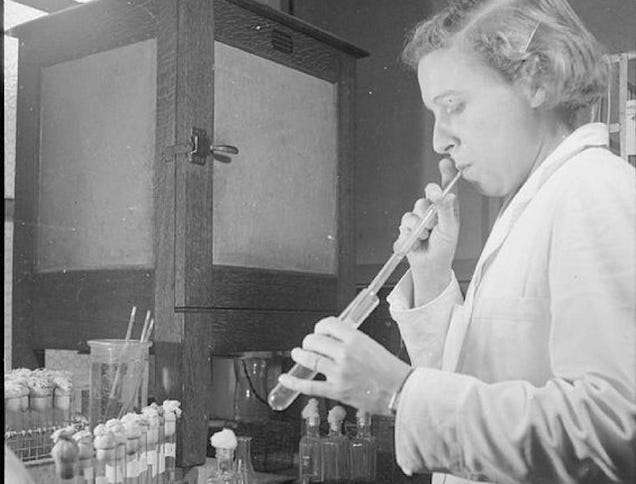
Here is the link for the article published in 1966:
http://www.dtic.mil/dtic/tr/fulltext/u2/640356.pdf
Friday, September 16, 2016
Could snail venom help treat diabetes?
The title may seem crazy, but this is an actual article from researchers out of Australia and the US. These scientists have figured out the 3D structure of venom insulin from a cone snail. The cone snail venom insulin is extremely efficient and acts much faster than human insulin. How could this help those facing diabetes though? Well, the efficiency of the cone snail venom insulin makes the cell signaling much faster and then increases how fast the insulin begins to take effect. They also discovered that the protein has the ability to bind to the receptors in human insulin, allowing the possibility for human applications. So the insulin from the cone snail is venomous towards fish, putting them in hyperglycaemic shock, but can be helpful for human diabetic therapies. The researchers on this project have already begun the process of finding ways to make better treatments with insulin that works much faster for diabetic patients. Why would it be advantageous to have fast acting insulin though? Most individuals with diabetes have to inject insulin before eating. Depending on the type of insulin, you can take it anywhere from an hour to 15 minutes before you eat. When you have to do an injection and then wait an hour before eating, this could cause problems in timing their meals. They could also eat too early or too late and would not have good control over their blood sugar levels. The faster the insulin works, then the closer to meal time the individual could take it, and would help maintain proper blood sugar levels. So, thanks to the creepy little snail shown below, we could eventually see improvement in diabetic therapies!

Here is the link if you want to read the full article:
https://www.sciencedaily.com/releases/2016/09/160912122604.htm
Here is the link if you want to read the full article:
https://www.sciencedaily.com/releases/2016/09/160912122604.htm
Friday, September 9, 2016
From scrapie to Bovine Spongiform Encephalopathy, or better known as Mad Cow Disease, prion diseases have interesting characteristics. According to the Centers for Disease Control and Prevention (CDC), a prion disease is a rare neurodegenerative disorder that is caused by "prions", which are pathogenic agents that can be transmitted and promote abnormal folding of proteins. These diseases can affect both humans and animals. One prion disease that I find to be very interesting is Fatal Familial Insomnia. This specific prion disease causes insomnia that gets worse and causes various symptoms to get worse, such as hyperventilation or the loss of control over your bodies movements, until the individual dies. This disease, like all prion diseases, has no cure. This is because the prion protein, which is caused by a mutation in the PRNP gene, is still a mystery when it comes to its' function. According to the National Center for Advancing Translational Sciences, scientists believe that the protein might play a key role in the brain since the mutation of the gene causes the protein to fold incorrectly and form clumps. These clumps will accumulate in the brain and destroy neurons, which leads to small holes in the brain. Researchers are actively working to find a cure, but unlike most viruses, prion diseases lack DNA or RNA. Prions are made of protein and a treatment or cure for this kind of disease has yet to be found.
Subscribe to:
Comments (Atom)